Topic: Foam nests in animals
Nests crop up everywhere, but one made out of foam? Might not sound like a great idea, but it is. And no surprise, it has evolved several times...
Life is full of hazards, not least to the young. So it comes as no surprise that many animals construct a nest of some sort.  One of the more intriguing examples is a nest made entirely out of foam, which holds eggs and sensitive juvenile stages. Such foam nests have a number of advantages, but foam is expensive to produce. To form bubbles in liquid, surface tension and high surface energy at the gas-liquid interface must be overcome, requiring considerable energy input. Foams are also difficult to maintain, because instability is an inherent property of fluid-based foams. Therefore, they tend to be uncommon in biological systems. Foam nests are, however, found in frogs, fish and some invertebrates.
Foam nests of tropical frogs
 Best known for their foam nests are various tropical and subtropical frogs of the Old and New World. The nests protect the eggs during development and are stable for several days or weeks. They allow eggs to be laid out of water, and many species suspend their nests in vegetation or construct them in underground burrows. Others build aquatic nests that float on the water surface.
Occurrence of foam nesting
Foam nesting has evolved independently several times in the frogs. The number of evolutionary origins depends on how one chooses to interpret frog systematics, a rather controversial issue. If we follow Julián Faivovich and colleagues (2012, Cladistics, vol. 1, p. 16),  then foam nesting has arisen in the American Leptodactylinae, the Afro-Asian Rhacophoridae, the Australian Limnodynastidae, the predominantly African Hyperoliidae, the Leiuperinae and the widely distributed Microhylidae and Hylidae. In the latter, the only foam-nesting species known to date is Scinax rizibilis, which is endemic to Brazil.
Interestingly, these independent evolutionary events seem to have taken different routes (as is so often the case with convergence). In the superfamily Hyloidea, foam nests have probably evolved from egg clutches laid in water, whereas in the superfamily Ranoidea, egg clutches laid on land formed the evolutionary basis for foam nesting. There have also been several independent transitions from foam nesting to eggs placed in a gelatinous matrix, for example in the leiuperine genus Pleurodema as well as in Rhacophoridae and Limnodynastidae.
Foam nest construction
 The first step in nest building is the production of a clear, thin, gelatinous substance in a modified part of the female’s oviduct. This structure has been referred to as a ‘foam gland’. This is, however, misleading as it is not actually a separate organ but rather an enlarged and folded portion of the oviduct’s posterior convolutions (so the term pars convoluta dilata (PCD) has been suggested). The females of all foam-nesting frogs seem to possess a PCD, although its size and form can vary, depending, for example, on the size and location of the foam nest. It is likely that the PCD itself has evolved several times. The secretion from the PCD traps air bubbles and can be beaten into a thick foam by limb movements when the male grasps the female in a form of pseudocopulation (known as amplexus). There is considerable variation between species in the particulars of nest construction. For example, male leptodactylines use their hind legs to whip the female’s cloacal fluid into foam, whereas in Australian limnodynastids the female herself creates the foam by “paddling” foreleg movements. Eggs and sperm are embedded into the nest, where fertilisation takes place.
Foam properties
Frog foams are amongst the most remarkable of biological materials, suggesting numerous potential technological and biomedical applications. On the one hand, they need to be stable and resilient against environmental challenges (especially microbial degradation), while on the other hand, they must not harm the delicate gametes and developing offspring. This poses a paradox, as foam production requires the activity of surface-active, detergent-like compounds, which typically damage unprotected cell membranes. However, frog foams do not rely on conventional small molecule surfactants, but employ a cocktail of special surfactant proteins and other molecules, which provides physical and biochemical stability whilst being compatible with the delicate gametes.
The biochemical properties of frog foam have been systematically studied in the American túngara frog (Engystomops pustulosus). Here, the foam shows remarkable mechanical stability, resisting compression and extension and not shearing easily, whilst being sufficiently elastic to form different shapes. Foam nests are produced in microbe-infested waters, where they remain intact for at least ten days, showing no degradation whatsoever. So what is this extraordinary foam made of? Mostly air, obviously, so the secret lies in the fluid phase, which consists of water and frog secretions. These secretions contain proteins and carbohydrates, mostly complex cross-linked mixtures of O- and N-glycans. 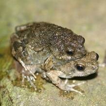 The protein content is surprisingly low, only 1-2 mg per ml – almost a hundred times less than that of foam a cook makes when whipping up an egg white! No lipids have been detected in túngara foam, which suggests an absence of conventional small molecule surfactant compounds. Still, túngara foam shows strong surfactant properties, and this is due to a remarkable set of proteins, the so-called ranaspumins. This is particularly intriguing, as the disruptive processes that presumably occur at the gas-liquid interface should lead to protein denaturation, so foaming of proteins is usually best avoided and proteins need to be specifically adapted for foam formation and stabilisation. In túngara foam, six major ranaspumins (RSN-1 to RSN-6) have been identified, all previously unknown. Their combination results in a stable, biocompatible and protective foam. The main surfactant seems to be RSN-2, which has an unusual amphiphilic amino acid sequence and lowers water surface tension rapidly and effectively, without infiltrating (and hence damaging) cell membranes. On its own, however, RSN-2 produces only short-lived foam. Long-term stability is achieved by aggregation and crosslinking of other foam components, such as carbohydrates, that results in a multi-layered surface structure. As RSN-3 to RSN-6 show carbohydrate-binding activity, they probably contribute to foam stabilisation. But they might have other functions as well. Their structure is reminiscent of that of lectins, sugar-binding proteins that occur in sensitive tissues of many animals and plants, where they counter microbial infections. RSN-3 to RSN-6 are therefore likely to provide defence, possibly not only against pathogens but also against parasites and predators. RSN-1 shows structural similarities to cystatins, a family of cysteine protease inhibitors, and might also be involved in antimicrobial defence. According to Alan Cooper and Malcolm Kennedy, “it is (…) intriguing to note that the combination of lectin and cystatin activities (…) is similar to that comprising part of the antimicrobial and anti-insect protection system of plant seeds, albeit achieved with unrelated proteins, representing an intriguing form of convergent evolution” (2012, Biophysical Chemistry, vol. 151, p. 102).
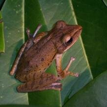 Recently, another unusual protein has been discovered in the Asian striped tree frog (Polypedates leucomystax). The structure of this blue protein, aptly named ranasmurfin, is also novel, showing no sequence similarity with identified and putative ranaspumins and containing a unique chromophoric crosslink. Ranasmurfin might help to stabilise the nest in the long term, provide camouflage or protect the eggs and juveniles from radiation. The detection of a very similar amino acid sequence in the archaeon Methanobrevibacter smithii has prompted the intriguing suggestion that frog ranasmurfin might have originated from a non-frog gene. The other components of P. leucomystax foam still need to be analysed in detail, but there seem to be some significant differences to túngara foam. For example, P. leucomystax foam fluid is sticky and syrupy, and the initial stability of the nest seems to depend on viscosity rather than surfactant activity. Overall, there seems to be great diversity in frog foam components. The foam nests of the northeastern pepper frog (Leptodactylus vastus) from Brazil, for example, contain another novel ranaspumin but apparently no antimicrobial defences.
Foam nest function
There is ample evidence that foam nests increase frog reproductive success, but what exactly is their advantage? 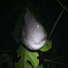 Several possible functions have been suggested, first and foremost protection of eggs and larvae from various environmental hazards, such as predation and microbial attack. Foam nests might also help to keep the eggs moist, preventing desiccation in a largely terrestrial environment. Temperature regulation could play a role as well. Another possible function is to increase oxygen supply to the eggs, hence speeding up their development. In the hylid Scinax rizibilis, for example, eggs with foam (and therefore more oxygen) indeed developed faster than eggs without foam (and less oxygen), and the large foam nests of the African rhacophorid Chiromantis xerampelina provide enough oxygen for all embryos until well after hatching. As the foam nests contain proteins and carbohydrates and might trap microorganisms, they can constitute a source of food for the developing tadpoles. This seems to be the case in Leptodactylus vastus, where the foam fluid also absorbs UV radiation and might thus prevent sun damage. It is generally likely that the nests serve more than one function in a particular species. In the túngara frog, for example, the nests not only provide protection from dehydration, predation and microbial attack but also accelerate offspring development by acting as ‘mini incubators’.
Foam nests of fish
More than 20 species of freshwater fish in three different families also construct foam nests.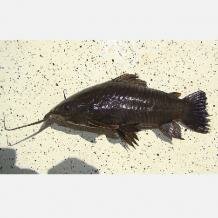 In the South American catfish Hoplosternum littorale, a floating foam nest is built and guarded by the territorial male. It consists of plant material that is held together by oxygen-rich foam. Nest construction starts with the fish swimming belly-up at the surface, whilst pumping water across the gills where it acquires mucus. The pelvic fins are then moved to mix water and mucus, trapping air bubbles that are broken into smaller and smaller bubbles. The resulting mass of foam is added to plant debris that the male carries up from the bottom with the help of a specialised pelvic fin spine. There is evidence that females also participate in foam making, but they are much less efficient than males and lack specialised pelvic fins. These catfish breed in tropical swamps, where the water is typically low in oxygen, and the floating nest probably helps to lift the eggs above the water surface, thus improving their oxygen supply whilst keeping them moist. Further possible nest functions might be related to protection from predators or temperature regulation. Other armoured catfishes in the subfamily Callichthyinae produce similar but less complex foam nests.
 The African pike (Hepsetus odoe) in the family Hepsetidae, which is found in tropical freshwater lakes, coastal rivers and swamps, also deposits its eggs in a foam nest. This nest is well hidden in dense vegetation and guarded by both parents. After hatching, the larvae employ a cement gland on the head to attach themselves to the nest, where they remain until later in development. Here, the main function of the foam nest seems to be predator avoidance, but it also serves as an adaptation to varying oxygen and water levels and might even provide a food source for the developing larvae. The foam-nesting habit is probably vital to the pike’s success in breeding in waters with large fluctuations in the annual flood cycle, where flood-dependent spawners cannot survive.
Foam nesting has also been documented in the family Anabantidae, for example in Ctenopoma damesi.
Biofoam production by tunicates
 The tunicate Pyura praeputialis is the only marine organism reported to date to employ biofoam in fertilisation. This free-spawning species inhabits the intertidal and shallow subtidal zone. In tunicate beds in Chile, masses of biofoam have been observed to form after release of eggs and sperm into the turbulent seawater. This biofoam had active surfactant properties and was seen to persist for several hours. Experiments have suggested that it serves to increase tunicate fertilisation success and retention of developing embryos and larvae in the rocky shore environment, thus allowing their effective settlement close to the spawning adults. In contrast to frogs and fishes, where the parents need to whip up the foam, foaming in these tunicates relies on wave energy. Hence, the parents only need to invest in production and release of foam precursors, making foam production less energetically costly.
Such biofoam production might be more common in intertidal marine organisms, as similar foam masses have occurred after mass spawning of sunstars Heliaster helianthus and chitons Acanthopleura echinata.
Biofoam production by insects
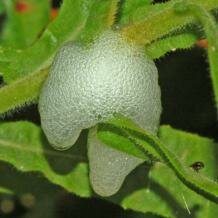 Amongst insects, foam production is probably best known from hemipterans such as spittlebugs. Here, the nymphs produce a froth commonly known as ‘cuckoo spit’, which surrounds them during development and provides protection from desiccation and possibly other environmental hazards. The froth seems to consist of a cluster of bubbles of different sizes, and its composition is complex but not well studied. It contains at least ten different polypeptides (mainly glycopeptides), acid proteoglycans, calcium and possibly more than one protein.
  The mineral composition is similar to that of the xylem sap ingested by these insects. Some other, less well-known instances of foam production occur in preying mantises and locusts, where females enclose their eggs in foam. In the former, the froth is produced by abdominal glands and hardens to form a protective capsule. In the latter, an intriguing function has been suggested for the foam. The desert locust (Schistocerca gregaria), one of the most feared pests, switches from a solitary to a gregarious phase, and this transition is under maternal control. Since the egg pod contains protective foam produced by accessory glands of the female, it has been speculated that this foam might include a pheromonal factor that affects offspring development and leads to gregarious individuals. Whilst this hypothesis was supported by some initial experiments, later studies failed to find evidence.
Cite this web page
Map of Life - "Foam nests in animals"
https://mapoflife.org/topics/topic_448_foam-nests-in-animals/
March 3, 2021

