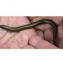
Topic: Viviparity in lizards, snakes and mammals
“In over 100 lineages of […] squamates, the oviduct has been recruited for viviparous gestation of the embryos, representing a degree of evolutionary convergence that is unparalleled in vertebrate history.” D. G. Blackburn (1998) Journal of Experimental Zoology, vol.282, p.560
Oviparity and viviparity
Forms of vertebrate reproduction can be divided into either ‘oviparous’ (egg-laying) or ‘viviparous’ (live-bearing), as first described by Aristotle in his Historia Animalium.  In egg-laying species, the developing embryo obtains nutrients from a store of yolk (termed ‘lecithotrophy’), whereas viviparity is a broad term encompassing varied divergence from simple egg retention in the female oviduct to elaboration of complex placental structures between fetal and maternal tissues. During viviparous gestation, embryos may acquire nutrients by digestion of ovum yolk (‘lecithotrophy’), intake of nutrients transferred from the mother (‘matrotrophy’), or – in certain viviparous amphibians and fish only – by consuming sibling ova or embryos in utero. Current evidence suggests that genetic and phenotypic changes essential to acquiring viviparity strongly (but not absolutely) resist reversal to oviparity. Accordingly, in this case of evolution, not only is the direction of the “tape of life” very much determined, but it is very difficult to rewind.
Viviparity in reptiles and other vertebrates
Viviparity has evolved more than 120 times in vertebrates, and surprisingly almost all of the transitions from oviparity to viviparity have occurred in the reptiles – the remaining occurrences being restricted to a few fish (sharks and rays), amphibians (certain salamanders and caecilians) and the well-known eutherian mammals, which are characterised by highly specialised placental structures for fetal nutrition. Over 85% of reptile species are oviparous, and yet viviparity has evolved convergently over 100 times. Most of these independent events have occurred in squamates (Squamata: lizards, snakes and amphisbaenians). Indeed, D. G. Blackburn notes that “in over 100 lineages of […] squamates, the oviduct has been recruited for viviparous gestation of the embryos, representing a degree of evolutionary convergence that is unparalleled in vertebrate history”.
Viviparity in squamates (lizards and snakes)
Factors affecting the evolution of squamate viviparity
Several explanations have been offered to explain the highly frequent transitions to viviparity within the Squamata, each revolving around unusual conditions that may act as preadaptations for viviparous gestation. One key feature is that, uniquely among the amniotes (reptiles, birds and mammals), squamate embryology features a membrane that extends from the embryo around the inner egg circumference, but before reaching the opposite side (termed the ‘abembryonic pole’), invades the yolk to form a thin ‘isolated yolk sac’. The outer part of the membrane (or ‘epithelium’, a single layer of cells) is then partly in contact with yolk mass and partly with the external egg surface or uterine epithelium, thus giving it the name ‘omphalopleure’ or yolk sac membrane.  Furthermore, the uterine epithelium of oviparous taxa is secretory to a limited degree, and the apposed omphalopleure epithelium is capable of uptake of water and ions, indicating a functional interaction that could also represent a preadaptation to viviparous gestation.
Squamate viviparity apparently evolved in response to both biotic and abiotic selection pressures for egg retention in the oviduct to successively later stages of embryonic development. The pattern of relationships between squamate taxa indicates that the genetic and phenotypic changes essential to acquiring such viviparity strongly resist reversal to oviparity. Accordingly, in this case of evolution, not only is the direction of the “tape of life” very much determined but it is very difficult to rewind. Due to the metabolic requirements of a growing embryo, so long as there is sufficient nutrient provision, selection for egg retention can only lead to viviparity when accompanied by parallel adaptations for increased gas exchange. Mechanisms to enhance respiration in utero include either eggshell thinning or its entire elimination, increased capillary blood supply (vascularisation) of the oviduct, increased extra-embryonic membrane vascularisation and surface area (e.g. by cell surface projections termed microvilli), and elevated oxygen-binding-affinity haemoglobin in fetal blood. Biotic factors aside, abiotic factors may exert significant selective force on novel reproductive strategies. In the case of squamate vivipary the importance of incubation temperature on embryo survival and offspring fitness has been shown in both cold and tropical environments, whereby the female, when her eggs are retained internally rather than laid in a nest, is able to control better the diurnal thermal regime experienced by the developing embryos.
Forms of squamate viviparity and placentation
Multiple forms of squamate viviparity and placentation have evolved, each involving modification of extra-embryonic and uterus membranes from an ancestral oviparous pattern. Most squamates reproduce by simple yolk-dependent (lecithotrophic) viviparity, featuring retention of large, yolk-rich eggs, eggshell reduction to a thin membrane, and limited exchange of nutrients for growth (histotrophy) between the uterine epithelium and isolated yolk mass omphalopleure. Accepting the definition of Mossman (1937) that a placenta is “any intimate apposition or fusion of the fetal organs to the maternal (or paternal) tissues for physiological exchange”, this arrangement has been termed an ‘omphaloplacenta’.  Gas exchange occurs across the well-vascularised interface between the outer-most embryo-derived membranes (the chorion and allantois) and the directly apposed uterine epithelium: this simple juxtaposition of fetal and maternal epithelia is technically termed a Type I or ‘epitheliochorial chorioallantoic’ placenta.
Moving on to consider other forms, some members of the Scincidae (e.g. Niveoscincus spp.) have a more specialised, Type II, placenta. Here the chorioallantoic placenta comprises a layer of large, cuboidal chorion cells closely associated with a ridged uterine epithelium of flattened (or ‘squamous’) cells, underlain by a dense capillary network. Yet more complex, Type III placentation is seen in a number of Australian skinks (e.g. Pseudomoia spp.) and the Mediterranean lizard Chalcides chalcides.  This morphotype is typified by differentiation of part of the chorioallantoic placenta into extensively folded, interdigitated uterine and chorionic epithelia. This structure is specialised for matrotrophic nutrient transfer, and is termed a ‘placentome’. Surrounding the placentome, gas exchange is facilitated by a ‘paraplacentome’ comprising microvilliated, enlarged chorionic epithelial cells apposed to a highly vascularised uterine epithelium.
 Finally, in the most advanced, Type IV placentation morphotype, famously found in the South American genus Mabuya, complex adaptations converge in intricate detail upon the derived placentation of eutherian mammals. Placental features shared between mabuyids and eutherians include a placentome of densely folded fetal-maternal epithelia, and a surrounding chorioallantoic placenta (paraplacentome) covered in clusters of cells (chorionic areolae) specialised to absorb products secreted from complementary uterine glands, and giant, binucleate chorion cells covered in microvilli. Adding to the list of remarkable convergences, both Mabuya and eutherians also ovulate minuscule (~1mm) yolk-free ova, provide >99% of embryonic nourishment by placentotrophy, and do so over a protracted gestation period (8-12 months).
Convergent evolution of placentation in squamates and eutherians
 Outside the amniotes, examples of simple and complex forms of vertebrate viviparity have been documented in the elasmobranchs (sharks and rays), osteichthyans (bony fish) and amphibians (anurans, salamanders and caecilians). However, none of these ‘anamniotes’ exhibit the extremely specialised placentotrophy as seen in Squamata and Eutheria. Although squamates and eutherian mammals are both amniotes, sharing a common extra-embryonic membrane arrangement with all other reptiles, birds and mammals, they are widely disparate taxa, having diverged from a common ancestor in the Carboniferous, >300Ma. This evolutionary separation, plus the occurrence of viviparity in Mesozoic marine reptiles (including ichthyosaurs, sauropterygians and mosasaurs) and an overwhelming number of squamate lineages, highlights the exemplary nature of reptilian viviparity as a case study in the power of convergent evolution to shape animal reproductive adaptations.
Cite this web page
Map of Life - "Viviparity in lizards, snakes and mammals"
https://mapoflife.org/topics/topic_331_viviparity-in-lizards-snakes-and-mammals/
March 4, 2021

