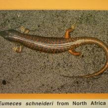
Topic: Mammal-like placentation in skinks (and fish)
“Only two types of vertebrates [have] evolved a reproductive pattern in which the chorioallantoic placenta provides the nutrients for fetal development. One is [...] the eutherian mammals […], and the other, a few lineages of the family Scincidae.” A.F. Flemming (2003) J Exp Zool 299A 33-47
 Most reptiles lay eggs (‘oviparous’ reproduction), within which the developing embryos obtain nutrients from a store of yolk (termed ‘lecithotrophy’). However, some 15-20% of reptile species retain their eggs internally and give birth to live offspring (‘viviparous’ reproduction). Of the viviparous species, most rely primarily on yolk for embryonic nutrition, but some lizards – and all of them are skinks – have evolved impressive adaptations that allow effective transfer of nutrients between mother and embryo (often referred to as maternal-fetal transfer, and termed ‘matrotrophy’). Skinks (family Scincidae) belong to the squamates, a group of reptiles encompassing lizards, snakes and amphisbaenians, and it is among skink species with complex, highly adapted placentas that we find the most astonishing similarities with the maternal provisioning mechanisms employed by eutherian mammals (and even certain fish). Similarity in placentation between these very distantly related groups – eutherian mammals and skinks – represents a shining example of convergent evolution, whereby selection for viviparous fetal nutrition has resulted in parallel and striking specialisations.
Placental structures in Mabuya and other skinks
South American skinks of the genus Mabuya (collectively called ‘mabuyids’) exhibit the most advanced placentation known in reptiles, and many of these placental features are shared with their close relatives in Africa, Mabuya ivensii and Eumecia anchietae. Distinctive features of mabuyid reproduction that are mirrored in the eutherian mammals include a long gestation period (8-12 months), ovulation of minuscule ova (<1000μm diam.), and provision of all embryonic nutrients via placental structures (termed ‘placentotrophy’).
 Nutrient transfer in mabuyids is achieved by means of a ‘placentome’, a region of highly vascular uterine wall which is densely folded and lined by thinned, vascularised chorion (outer embryo-derived layer). This dense mass of projections resembles the tree-like (‘deciduate’) placentae of artiodactyl mammals; in particular, the Mabuya heathi placentome is convex in shape, exactly like that of bovid and cervids. The mabuyid placentome also converges on the mammalian form in that both exchange structures allow for transfer of lipid molecules.
  A ‘paraplacentome’ region of the chorion (along with the underlying allantois) surrounds the placentome and is covered in clusters of cells termed the ‘chorionic areolae‘. Areolar cells are enlarged and covered in finger-like protrusions (microvilli) specialised to absorb products secreted from the uterine glands that overlie each of them; similiarly functioning areolae are also present in the mammals, specifically carnivore and primate placental tissue. Non-areolar chorion cells are adapted for respiration and nutrient exchange, specifically by adaptiations that serve to increase the surface area and decrease the diffusion distance between fetal and maternal tissues. The chorion is also modified by forming a thinner than usual layer (epithelium) of enlarged, binucleate and villiated cells.
  Remarkably, these resemble the situation in ungulates and several other mammal groups.
Skinks with slightly less extreme placentation than Mabuya and Eumecia provide further illustrations of convergent evolution. For example, after implantation of a mammalian embryo the lining of the uterus forms domed structures; identical ‘uterodomes’ are found in the skinks Niveoscincus metallicus and Eulamprus tympanus. These are two species in which a specialised placentome forms, but it is less well developed than in the mabuyids. Furthermore, in another skink Pseudemoia entrecasteuxi, specialised chorion cells of the paraplacentome region uniquely resemble those of the chorionic epithelium in the eutherian mammal species Scalopus americanus (American mole). These points are especially notable as the species mentioned are unrelated to Mabuya, and yet the path of evolution has brought all of them to placental adaptations that are clearly shared with mammals.
Placental structures in fish
Fish possess extra-embryonic membranes unlike those of skinks and eutherian mammals, and yet a certain degree of convergence in placental adaptations also exists between these three groups.  Viviparity has evolved in fish approxiately 30 times, occurring in the actinopterygians (ray-finned fish), sarcopterygians (lobe-finned fish/coelocanths) and chondrichthyes (cartilaginous fish, e.g. sharks, rays and skates). Embryonic nutrition within the follicle/ovarian cavity or uterus is enabled by a diversity of both aplacental and placental means. Among the placental adaptations, the main parallels with skinks (and mammals) include: increased vascularisation, surface area and thinning of the uterus wall, microvilliation of surfaces for respiration and/or nutrient absorption, and development of various placental analogues. Specialised placental structures in fish include projections of the uterine wall termed ‘trophonemata’, intestinal projections termed ‘trophoteniae’, ‘yolk sac placentae’ (formed after yolk depletion and similar to the marsupial placenta), and ‘follicular pseudoplacentae’ in which a direct blood vessel links fetal and maternal circulatory systems. Trophonemata (found in rays) converge most closely with a skink-like mode of placentotrophy, although the yolk-sac placenta also mimics a form of placentation seen in some less specialised squamates.
Cite this web page
Map of Life - "Mammal-like placentation in skinks (and fish)"
https://mapoflife.org/topics/topic_336_mammal-like-placentation-in-skinks-and-fish/
April 22, 2021

